Ecografía 6D

GET TO KNOW YOUR CHILD FROM THE VERY FIRST DAY WITH THE MOST HIGH-TECH PRENATAL DIAGNOSIS

GENERAL ELECTRIC VOLUSON E10 BT18

Clinica Ripoll has just acquired a General Electric ultrasound machine: the Voluson E10 BT18, with the most advanced technology in 2D prenatal testing, Doppler color, e-stic and 5D, 6D or live HD Silhouette ultrasound. The Voluson ensures the highest quality, speed and flexibility for ultrasounds. It is currently the most advanced version for ultrasound, and Clinica Ripoll’s latest acquisition.

IT IS THE FIRST EXISTING ULTRASOUND SYSTEM OF THESE CHARACTERISTICS IN BARCELONA

Clinica Ripoll is still spearheading the integration of the best technology, which makes it an important referent in Barcelona. Particularly, the Department of Prenatal Testing lead by Dr. Guillermo Ripoll has been further improved with the acquisition of this machine.

The GE Voluson E10 BT18 is able to show all of the baby’s details, and it is considered the present and the future of prenatal care: it offers real time pictures with deeper penetration and better resolution, which allows for more precision in observation and detection of any complications there might be from the very first weeks of pregnancy.

It is true that the GE Voluson E10 BT18 is capable of early observation of important areas such as the brain and heart (veins, arteries) of a fetus, but it can also aid in a wide variety of complex female-related examinations thanks to 3D/4D/5D-HD live and 6D high-definition ultrasound for the detection of deformities in the uterus (septum, bicornuate uterus…) among others.

This new instrument is of great utility both for patients and professionals. Future parents have the opportunity to see, with much better precision and clarity, what their child is going to be, and they may also have an early and more certain idea of how the child’s health will develop. Moreover, in Clinica Ripoll we count on the most powerful existing tool currently available in the market that allows us to work more efficiently and give better advice in the areas of prenatal and reproductive health.

Clinica Ripoll is always seeking to integrate the best technology, because we are aware that the best and most successful outcomes are the result of combining medical knowledge with top-notch technology.

Clinica Ripoll’s Department of Prenatal Testing has become a reference in its area of expertise by offering a comprehensive service in the early diagnosis of fetal diseases during pregnancy. It is in fact a role model for many gynecologist colleagues in Barcelona.

In general, pregnancy monitoring and caring for mothers is performed by our obstetrics-specialized gynecologist, and the women are likewise put in the care of Clinica Ripoll’s Department of Fetal Medicine for specific monitoring of the baby. There are different key moments for verifying the proper development of the baby, in order to ensure that it does not suffer from any genetic disease and to prevent any complications during birth.

In case of a severe fetal pathology, patients are put in the care of Level III Hospitals for medical treatment in specialized units and/or fetal surgery for specific cases (for instance, feto-fetal transfusion for monochorionic twin pregnancies, spina bifida, etc.)

We offer and perform all currently available testing in the area of fetal medicine with the aid of highly-qualified specialists and top-notch avant-garde technology.

We offer:

  • Ecografical biochemical aneuploidy screening programs
  • Non-invasive prenatal testing during the 1st trimester (trisoNIM test)
  • Chorion biopsy
  • Early and/or late amniocentesis
  • Genetic testing (aneuploidy – fetal karyotype screening)
  • Molecular karyotype (karyoNIM test)
  • Pre-eclampsia screening
  • High Definition ultrasounds
  • Doppler color ultrasounds
  • 3D/4D Ultrasounds and the innovating HD Live Silhouette and HD Live Studio ultrasounds

The director responsible of the Department is Dr. Guillermo Ripoll, a gynecologist specialized in prenatal testing and fetal medicine with dedication and experience in this area. He belongs to the ISUOG based in London.

In Clinica Ripoll, we know that for you to enjoy your pregnancy, its proper monitoring must be carried out by specialists. Follow all and any advice given by your doctor so you can rest assured during this whole process. In Clinica Ripoll, we offer the medical counseling you need in order to feel safe and accompanied during the most important moments of your pregnancy. A trustworthy surveillance and monitoring of your baby’s development is essential to experience a calm, healthy pregnancy.

Any OB-GYN in charge of a patient can ask for any necessary prenatal testing according to his or her pregnancy monitoring plan, or the patient herself may ask to attend a prenatal testing consultation at Clinica Ripoll.

We summon the patient and explain to her what each test to be performed is about, with the proper medical and genetic advice for each particular case in order to help her make decisions and for her to always be fully accompanied. In addition, we create a comprehensive report of each test and ultrasound that is then delivered to the patient.

Prenatal monitoring of the fetus during pregnancy can be divided in three different stages corresponding to the three trimesters of the gestation of a human baby.

FIRST TRIMESTER

  1. Ultrasound assessment between the 6th and 9th weeks.

During the first trimester, it is of the utmost importance to ensure fetal viability, in case of singleton or multiple pregnancy (dichorionic or monochorionic, commonly known as twin pregnancies), as well as a correct implantation and the detection of certain chief pathologies such as hematomas and/or other upcoming complications.

  1. Ultrasound assessment – 12-week gestation – EBA-Screening

The ecografical biochemical aneuploidy screening consists of the integration of multiple data resulting from the ultrasound performed after 12 weeks of gestation and a combined biochemical screening analysis of chromosomal alterations (e.g. Down syndrome, Edwards syndrome, Patau syndrome, etc.).

The ecografical biochemical aneuploidy screening is the current screening alternative with the highest detection rate (95-99%), much higher than that of traditional triple biochemical screening (65). Its false-positive rate (tests yielding results of augmented risks of chromosomal pathology, but with children born healthy) is only 5%. We have been performing this test in our facilities since 2003 and, with over a thousand documented cases, our false-negative rate (children born healthy after regular testing) has been 0%. Once established as the monitoring method during the first trimester, this alternative allows for a highly trustworthy and precise estimation of the actual risk of fetal chromosomal disorders.

Methodology

Step 1: Blood testing of the future mother between the 10th and 12th weeks of gestation. The values of two chemical parameters will be specifically relevant: free Beta-HCG (pregnancy hormone) and the PAPP-A protein (originating the placenta).

Step 2: Transvaginal-abdominal high definition ultrasound between weeks 11 and 13.6 of gestation. It will be performed according to the criteria of the Fetal Medicine Foundation to get the values of the Ecografical Biochemical Aneuploidy Screening program (measurements of nuchal translucency (NT), the presence of the nasal bone and of the ductus venosus, and many other ultrasound parameters). Additionally, during the ultrasound of 12-week pregnancies, an anatomic fetal assessment is carried out so as to rule out certain major structural malformations such as anencephaly and omphalocele observable at this stage of gestation. We also examine all the developing fetal anatomy: brain, face, mouth, eyes, lungs, liver, kidneys, hands, feet, heart… For some cases, we can even determine the sex of the fetus.

To make the specific calculations, we use a risk calculation software (from the Fetal Medicine Foundation: www.fetalmedicine.org ) to determine the possible chromosomal risks of the fetus (based on a rate obtained from the results of the biochemical analysis alongside the values taken from the ultrasound and the age of the mother). Our results are audited annually by the Fetal Medicine Foundation in order to guarantee the top performance in our endeavors involving chromosomal alterations detection.

The result is shown in the form of a fraction (with a varying limit value according to the age of the patient). Depending on the result, and the personal and familiar precedents in each case, other techniques might be recommended to deepen the analysis and rule out fetal pathologies shown after an incorrect diagnosis. A non-invasive prenatal test (trisoNIM test) could be performed as well as a Chorion biopsy or an amniocentesis.

  1. Non-invasive prenatal testing (trisoNIM test) during first trimester between the 10th and 13th weeks of gestation

Non-invasive prenatal test for the detection of alterations in chromosomes X, Y, 21, 18, 13 which also allows for the identification of the fetal sex.

Variation in a fetus’ number of chromosomes signals the presence of complex genetic diseases. They may occur after an increase in the number of chromosomes (e.g. Trisomies: Down syndrome) or for a lack of chromosomes (e.g. Monosomies: Turner syndrome). Such alterations are rather frequent in human beings, and the objective of Prenatal Diagnosis is to detect them early on so as to inform the parents of all the details involving the consequences of these diseases and the prospect and impact they may have.

Since 2013, we have been applying the non-invasive genetic analysis known as trisoNIM test, which analyses the number of chromosomes Y, X, 21, 18 and 13 by measuring the relative amount of fetal DNA in the mother’s blood. That is, a simple blood test on the mother can determine the chromosomal proportion of the previously mentioned chromosomes in the fetus. Furthermore, analyzing the sexual chromosomes (X and Y) can accurately pin-point the sex of the fetus. The specific assessment of these 5 chromosomes is undertaken because they are responsible for 95% of all chromosomal alterations detectable by prenatal diagnosis.

The trisoNIM test can be performed after the 10th week of gestation, for singleton pregnancies (including cases of egg donation) or twin pregnancies of two fetuses (except cases of egg donation). It should be noted that for twin pregnancies only alterations in chromosomes 21, 18 and 13 are analyzed (alterations in chromosomes X and Y cannot be analyzed, so the sex of the babies cannot be identified in this type of gestation)

It is important to underline the high reliability of the results obtained through the trisoNIM test, which is based on the latest advancements in non-invasive prenatal testing, with a detection rate of >99% for the analyzed chromosomes and a fake-positive rate of less than 0.1%. The great improvement that trisoNIM provides is that there is no risk for the mother nor the fetus since it is a simple extraction of a blood sample from the mother, and the results are given in a week.

SECOND TRIMESTER

  1. Comprehensive chromosomal analysis

There are two different invasive methods for the collection of fetal material for chromosomal analysis (chorion tissue and amniotic fluid).

Indications for the application of an invasive test:

  • EBA-Screening with a risk higher than the mother’s age
  • Altered trisoNIM test
  • Mother older than 37 years old
  • Previous offspring or miscarriage with a chromosomal alteration
  • Previous offspring with structural alteration possibly transmitted through genes
  • Family history of monogenic disease(s) (hemophilia, cystic fibrosis, etc.)
  • Family history of altered karyotype
  • Willingness to become mother
  • Suggestive ultrasound signs of chromosomal malformations (nuchal edema, omphalocele, etc.)

The two most common methods are:

  1. Chorion biopsy – 13 weeks of gestation

It is performed transvaginally between the 12th and 13th weeks (or transabdominally with local anesthesia) with ultrasound. The rate of post-puncture fetal loss is lower than 1 out of every 200 in expert hands. It is especially useful when a very early diagnosis is required to rule out any risk of chromosomal alteration, since the results are given in 7 days. It is the most popular choice for the majority of genetic diseases such as hemophilia and when EBA-Screening yields an altered result.

  1. Amniocentesis – 16 weeks of gestation

It is performed transabdominally after 15 or 16 weeks with ultrasound. The post-puncture fetal loss rate is lower than 1 out of every 200 in expert hands. Most indications come from fetal structural alterations or maternal old age, or precedents showing genetic risks.

When amniotic fluid is collected, two assessments may be performed:

Fetal karyotype: It entails the determination of the genetic map of fetal cells located within amniotic fluid in order to rule out the existence of a chromosomal disease. The regular expected outcome would be 46XX in the case of a girl and 46XY in the case of a boy. Normally, the results are given 2-3 weeks after cultivating the cells, although the possibility of obtaining a part of the results after 3 days through QF-PCR testing exists.
Molecular karyotype or CGH array: Comparative Genomic Hybridization is the most advanced method of genetic analysis that allows for a comprehensive examination of genomes so as to detect the increase or loss of genetic material. It presents a higher resolution than conventional karyotype, detecting more than 128 genetic syndromes that could not be detected otherwise and that cause malformations and/or mental retardation in varying degrees of impact. The results are given after 5-7 days along with a thorough and clear report referencing each of the diseases considered for analysis. It is especially prescribed for cases of normal karyotypes showing ultrasonic alterations, repeated miscarriages and fetuses with structural chromosomal anomalies.

  1. 2D High-resolution morphology ultrasound ( 20-22 weeks of gestation )

It is performed after 2 weeks with the use of high-resolution technology. Known as “high-resolution ultrasound”, it implies the systematic examination of each aspect of fetal anatomy in search of structural alterations. After 20 weeks of gestation, the fetus already has all fully-formed organs, measures between 14 and 16 cm (roughly 5.5 – 6.2 inches), and weighs around 260 grams (9.17 oz.). It is then possible to undertake the systematic examination of all organs and fetal structures, looking for different alterations. Moreover, certain markers of chromosomal alteration are evaluated such as nuchal edema, hydronephrosis (short femur), and brain anatomy, among others.

A detailed assessment of the fetus’ heart is performed in order to detect congenital abnormalities. This guarantees the proper planning of the pregnancy period and of the delivery in a level III hospital in case of severe heart disease.

Thanks to Doppler color flow imaging, the resistance indexes of uterine arteries can be determined so as to define the risk factors of pre-eclampsia (hypertension during pregnancy) and delayed intrauterine growth.

It is evident, then, that this ultrasound is of the utmost importance, since both the specialist and the future parents can observe the entire fetal anatomy, which is a relief when no fetal malformation is detected.

As a novelty coming with this ultrasound, one should ask for a 6D ultrasound to see the face of the fetus, so that any possible syndromes can be detected. In addition, future parents can create an early bond with their offspring by watching with great clarity his or her face.

  1. 3D/4D/5D-HD live ultrasound and a novelty: 6D High-resolution (28-30 weeks of gestation) )

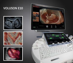
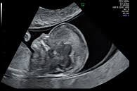
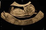
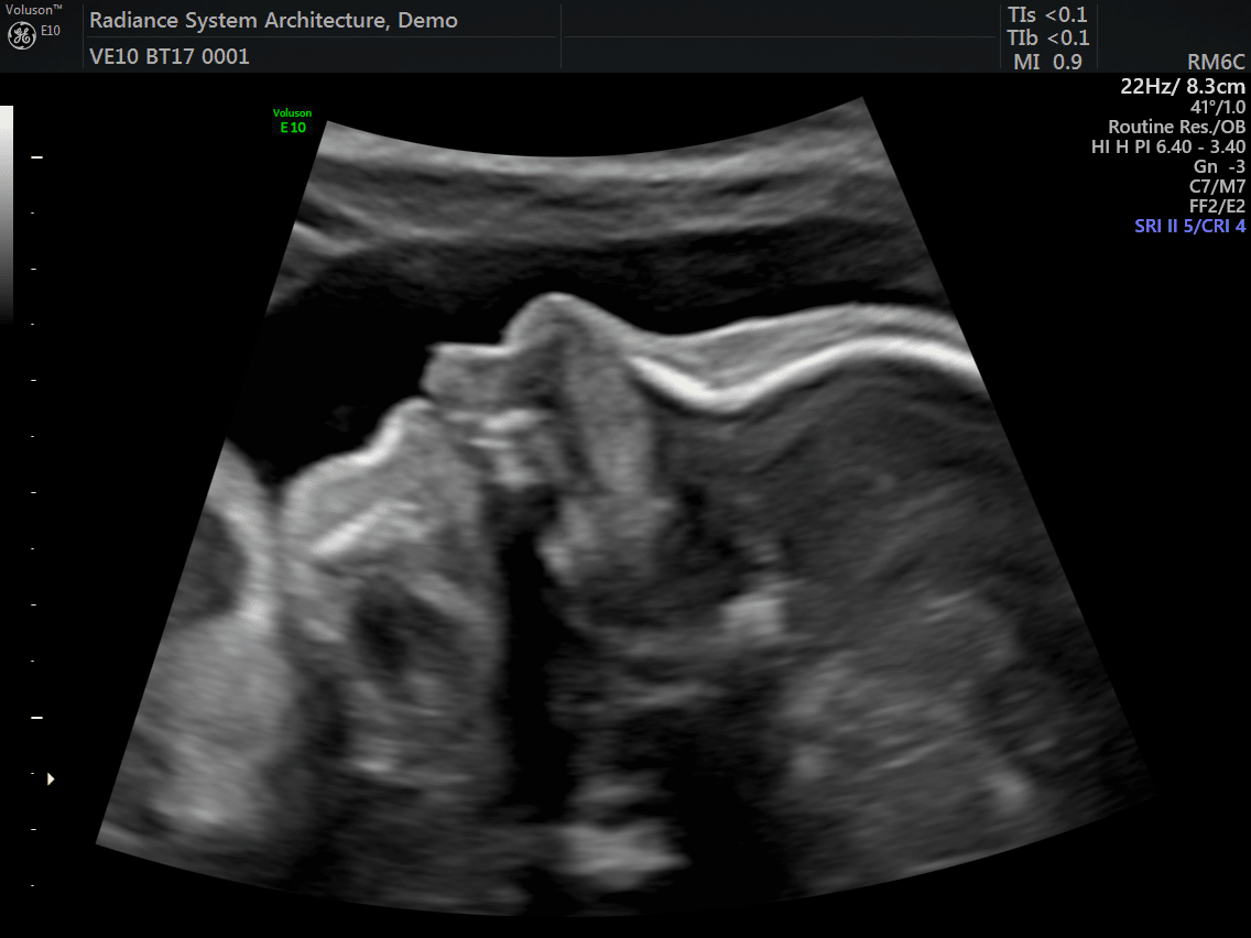
Being one of the most recent and impactful innovations in the domain of prenatal diagnosis, 5D HD Live Silhouette and HD Live Studio ultrasounds are recommended in a routine application for gestation monitoring. Two aspects should be underlined: On one hand, it provides real-time 3D images instead of flat 2D images, contributing to the accuracy of the diagnosis; on the other hand, it has been demonstrated that such images further strengthen the bond between mother and child even before delivery.

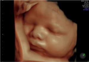
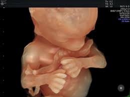
It is particularly remarkable, with regards to 5D HD Live Silhouette and HD Live Studio ultrasounds, that the resulting images allow parents to establish a strong bond with their offspring. Parents actually see and understand what their offspring looks like. This strong bond has been proven by psychological research involving the impact before and after 2D, 5D HD Live Silhouette and HD Live Studio ultrasounds..

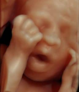
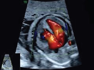
In certain cases, where a fetal pathology exists, 5D HD Live Silhouette and HD Live Studio ultrasounds are particularly useful for the explanation of some malformations, such as facial ones, since the parents are able to watch the alteration in a clear and understandable way. The possibility of using the 3 planes of space helps to complement the malformation diagnosis and provide better detection of otherwise non diagnosable alterations. This aspect is indeed distinctively important from the point of view of the concrete malformation diagnosis of the baby and his or her future quality of life.

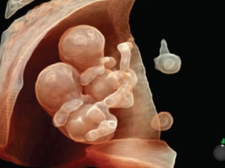
Additionally, a number of European studies show that the use of 5D HD Silhouette and HD Live Studio ultrasounds has enabled a deeper understanding of the development of fetal movement and behavior, previously unknown. It has been discovered that fetuses may open their eyes after 18 weeks. It has also been observed that the little ones laugh, cry, scratch and use their mouths in a sucking fashion after 26 weeks of gestation. All these complex functions require vital neurological development.

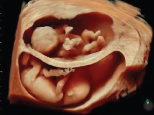
5D HD Live Silhouette and HD Live Studio ultrasound is a powerful working tool granting a deeper understanding and interpretation of human behavior during the fetal stage. The clarity and high resolution of the images further improves the capacity of the abnormality diagnosis. It allows us to have an idea of what the child is going to be like before his or her birth.

THIRD TRIMESTER

Biometrics – ultrasound after 32-34 weeks of gestation

It is performed between the 32nd and 36th weeks of gestation by implementing high-resolution technology. Fetal anatomy is once more analyzed in search of abnormalities that might appear during the third trimester, such as bowel obstructions, skeletal dysplasia, late brain alterations (such as ventriculomegaly), etc.

By undertaking fetus biometrics, it is possible to determine the state of fetal growth and to know the risk of delayed intrauterine growth, of low weight in relation with gestational age or of macrosomia.

It is during this time that the placenta must be evaluated regarding the ultrasonic aspect and position so as to plan ahead before delivery. The estimated weight of the baby at birth and his or her position in the womb are important in the adoption of preventive obstetric measures for each case.

According to the results shown by biometrics, it might be necessary to perform a fetal hemodynamic assessment using Doppler color in the womb, placenta and fetus.

For particular cases, it may be required to perform other types of special ultrasound, for instance:

  1. Echocardiography: Anatomic and functional assessment of the fetus’ heart. It is used in real time as with Doppler in its various modes.

Indications:

  • Precedents of congenital heart abnormalities
  • Diabetes Mellitus
  • Epilepsy under treatment
  • Augmented pathological nuchal translucency (Nuchal fold)
  1. Fetal Hemodynamics

Where slow fetal growth is suspected, it is necessary to evaluate the well-being of the fetus through Doppler coloring in fetal, placental and uterine blood vessels. A biophysical fetal profile must also be performed including fetal tone and respiratory movements. The idea is to complement the cardiotocographic register of the fetal situational assessment in order to ensure the proper planning of the delivery.

  1. Cervical Ultrasound

In case of precedents of premature delivery or incompetent cervix, or with patients having cervical cerclage, it is necessary to inspect the cervix by measuring the length of the cervical canal and see if there is any dilatation and/or funnel. This way, the risk of preterm birth can be determined, and the proper measures concerning the treatment of tocolysis and prophylactic cerclage can be established.